
How a team at IIT Madras built the world’s most detailed atlas of the human brainstem — and why it matters
For more than a hundred years, neuroscientists have studied the human brain the way early explorers charted unknown continents: assembling a vast picture from scattered glimpses. The problem is that the terrain is enormous. The human brain holds roughly 86 billion neurons, yet a pathologist diagnosing a disorder like Alzheimer’s typically examines only a handful of tissue slices. Most of the map stays blank.
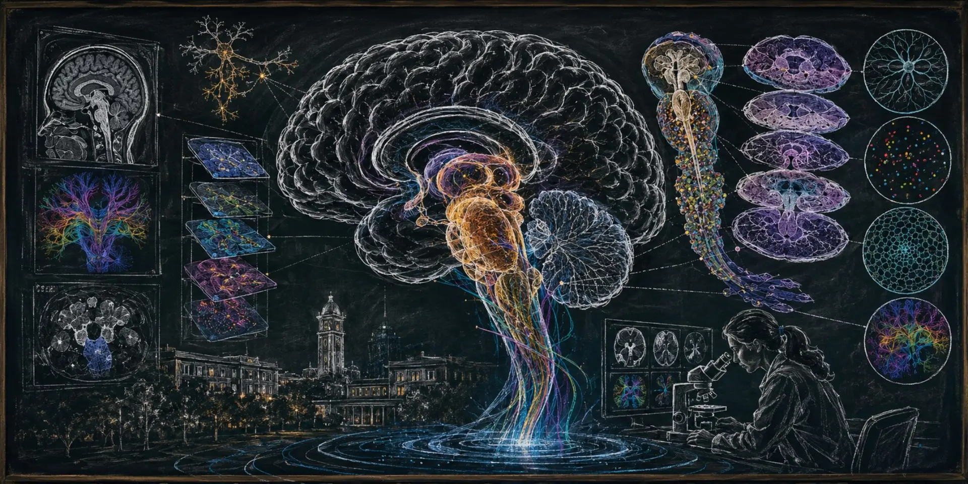
A team of scientists in India believes it has taken a real step toward closing that gap. Researchers at the Sudha Gopalakrishnan Brain Centre (SGBC) at the Indian Institute of Technology, Madras have produced what they call the world’s most detailed three-dimensional atlas of the human brainstem at cellular resolution — a digital map that lets a scientist zoom seamlessly from an MRI scan of the whole brain all the way down to a single nerve cell.
What they built
The atlas is called ANCHOR, short for Atlas of Neurochemical Characterization of the Human Brainstem with 3D Reconstruction. It stitches together more than 500 tissue sections drawn from fetal, childhood and adult brains. Rather than leaning on expensive molecular techniques, the team built the map from high-resolution microscope images, identifying more than 200 clusters of brain cells and nerve pathways. This is typical of India’s frugal mindset to produce innovations. Eight chemical markers help tell one cell type from another, producing one of the clearest pictures yet of a region that has long resisted close study.
A sliver that keeps you alive
The brainstem takes up only a small fraction of the brain, but it does some of the most essential work. It connects the brain to the spinal cord and governs breathing, heartbeat, sleep, wakefulness and movement. Damage to even a tiny cluster of cells there can be catastrophic. The trouble is that the region is packed so densely that mapping it in detail has frustrated scientists for generations.
Bridging two separate worlds
ANCHOR’s real breakthrough is not just another anatomical chart. It links two fields that have mostly operated apart: medical imaging, which shows the brain as a whole, and cellular pathology, which reveals it one cell at a time. Shubha Tole, a neuroscientist at the Tata Institute of Fundamental Research, called the project an “unprecedented integration” of engineering, neuroscience and medicine, and said it “puts India at the international table.” The gap it addresses is old and stubborn. Doctors still begin much as they did a century ago, when Spanish neuroscientist Santiago Ramón y Cajal pioneered the field. “As a neuropathologist, I begin by examining an entire brain with the naked eye before looking at small pieces under the microscope,” says Rebecca Folkerth, affiliated with Harvard Medical School and New York University, who collaborated with the SGBC team. “For Alzheimer’s disease, we may examine only 15 to 20 sections — just a fraction of a percent of the whole organ.” Modern MRI shows the entire brain but misses the cells; the microscope shows the cells but only in isolated slivers. ANCHOR
effectively zooms in nearly a thousandfold, from the millimeter scale to the micron scale, and holds both views in one place.
The bigger picture
The atlas was unveiled at the 3rd BRICS Neuroscience Symposium held at IIT Madras in June 2026, and the team has made it freely available online — open to scientists, clinicians and patients anywhere. It is also just the first installment. SGBC, which earlier released DHARANI, a 3D atlas of the developing fetal brain, envisions building cell-resolution maps across the entire human lifespan and the full range of brain disease.
For readers in the United States and worldwide the payoff is practical. The resource costs nothing to use, and sharper maps of the brainstem could eventually mean better diagnosis of the disorders that quietly begin there. A century after Cajal, much of the brain’s landscape is finally coming into view.

